Health Sciences Education and Pathology Resources and Facilities
Resources & Facilities Heading link
Health Sciences faculty are located on the UICOMP campus in the B2 wing. Also located on campus are the Anatomy Tech Lab, Anatomy and Pathology Gross labs and the Pathology / Histology Microscope lab.
Anatomy Labs Heading link
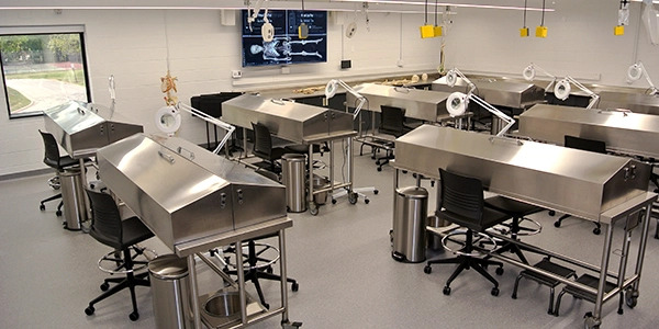
Gross Anatomy Lab
A state-of-the-art lab used for dissection of human cadavers*.
The lab is outfitted with:
- eight dissection tables
- suspended and portable lighting
- variety of anatomical models
- Additionally, the lab is equipped with a high resolution monitor that is integrated with mobile technology platforms such as iPad Pros and Apple TV. The iPad Pros are used by the instructors and students to film and photograph dissections for further review of anatomy content.
*Cadaveric dissection at the University of Illinois College of Medicine Peoria is possible only through the generous, whole body donations made by those individual donors to the Anatomical Gift Association of Illinois (AGA). For more information regarding whole body donation, please contact the AGA. While UICOMP does not directly take bodies for donation, the AGA accepts donor requests to be directed to a specific institution, such as UICOMP.
Anatomical Gift Association of Illinois (AGA)
312-733-5283
info@agaillinois.org
Technology-Assisted Anatomy Lab Heading link
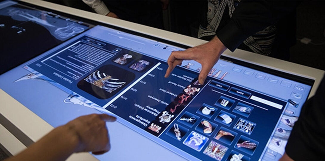
Anatomy Lab Heading link
The Technology-Assisted Anatomy Lab houses the Anatomage table, a high resolution virtual dissection table. The Anatomage table is a fully interactive life-sized touchscreen imaging system in table form that displays detailed human anatomy systems and structures. Users are able to:
- explore and dissect the human body in different planes and sizes,
- review real case studies,
- view digital histology slides, and
- observe radiological imaging all with the touch of a finger.
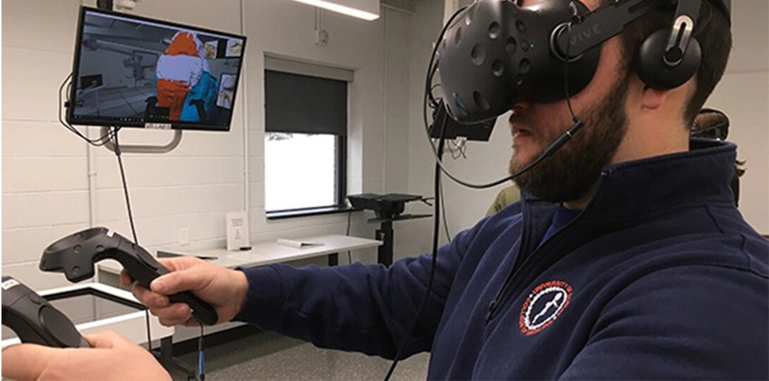
The lab also contains:
- a mobile ultrasound unit,
- two treatment tables for students to learn and practice surface anatomy and ultrasound imagining skills,
- a multimedia computer station furnished with interactive anatomy software, and
- VR/AR stations utilizing the HTC Vive technology.
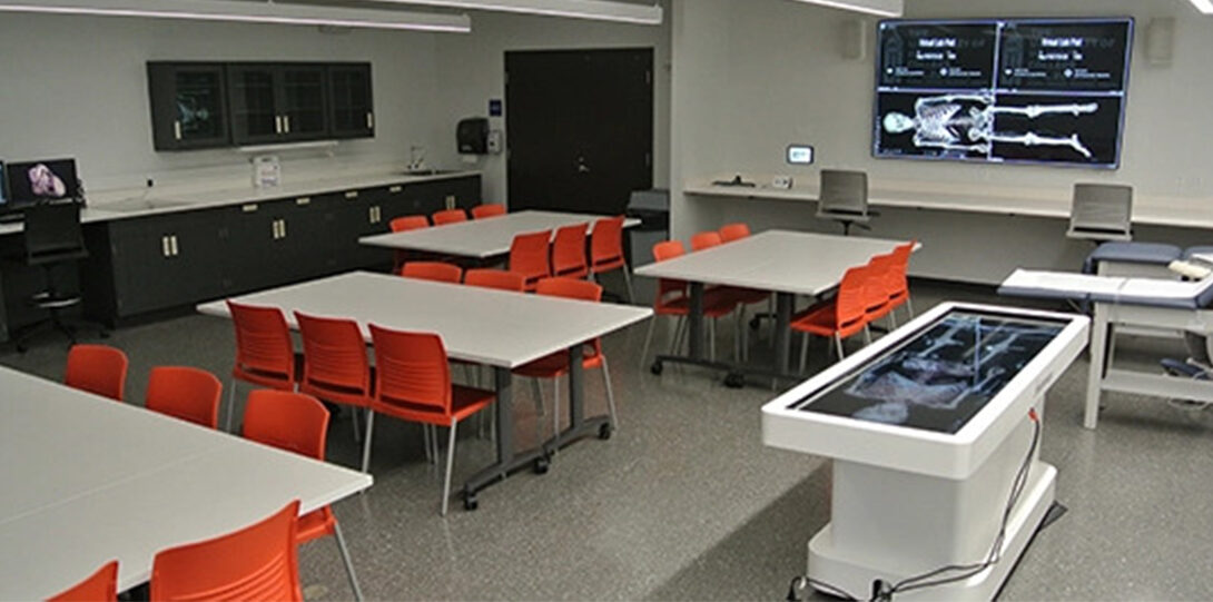
- Like the adjacent gross anatomy lab, the technology-assisted anatomy lab also boasts a large, high resolution monitor which can display up to four different inputs simultaneously, including input from the Anatomage table, the ultrasound machine, and the iPads.
The monitors in both lab spaces are integrated with one another, allowing the learning activities viewed on one lab’s monitor to be mirrored for viewing by students on the monitor in the adjacent lab. This provides for a synchronized learning environment between two separate lab spaces.
Lastly, a sophisticated negative return ventilation system ensures the quality of air in both lab spaces to enhance the overall learning environment.
Pathology / Histology Labs Heading link
Microscope Lab
While faculty are using Virtual Microscopy and digital images for parts of the curriculum, the Peoria campus also enjoys the use of a large collection of glass slides for use in pathology and histology studies. Students have the opportunity to explore glass slides demonstrating normal histology, pathologic processes and a variety of infectious diseases (such as malaria smears) using a 5 headed microscope. In laboratory sessions this allows students and faculty to examine slides together and discuss the microscopic findings. Located in the B wing, students have an opportunity to learn and use microscopes to view
Gross Pathology Lab
The Peoria campus has an impressive collection of gross pathology specimens which are incorporated into various pathology laboratory sessions. Students can appreciate first-hand the effects of various disease processes on targeted organs.